
Laser Treatment to Superficial Cornea in Midperiphery
Corneal Topographic Changes Direct Light to Multiple Areas Outside of Fovea
If an Image Falls on the Preferred Retinal Loci Vision Can be Enhanced
Case
An 82-year-old male with age-related macular degeneration was referred to Bochner for consideration of a new Cornea Laser procedure. Best-corrected acuity was 20/125- in his right eye and 20/125 in his left eye. He was pseudophakic in both eyes and had well positioned posterior chamber implants. Fundus examination revealed geographic macular degeneration in each eye.
Computerized topography was normal without evidence of irregular astigmatism. Vision testing at near with the GM Potential Acuity Charts showed better vision at near compared to distance by 4 lines in each eye. This indicated that the patient had functioning retinal cells in the macula outside of the fovea, and the potential for enhanced vision with this Corneal Laser procedure. OCT imaging showed no evidence of a wet macula.
What are the Surgical Options to Improve Vision?
- Intraocular Minature Telescope
- Corneal Laser for Macular Degeneration
The Intraocular Telescope is a more invasive procedure that involves implanting a specialized telescope in to the capsular bag. This procedure can magnify the image on the fovea to enhance vision but reduces the field of vision.
The Corneal Laser procedure is relatively noninvasive, very easy for patients to undergo, and has an excellent prognosis with some functioning retinal cells in the macula. The procedure involves the use of a specialized corneal laser with a proprietary wavelength.The laser creates four focal spots on the cornea at the level of anterior storm in the midperiphery to redirect light on to functional retinal areas. No postoperative medication is required.
What was Done?
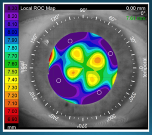
The Corneal Laser procedure was performed to both eyes in the operating room. The patient was comfortable during and after the procedure. Slitlamp examination immediately after the procedure revealed 4 focal white opacities at the level of the anterior stroma approximately 0.5 mm in diameter.
No postoperative drops were required. Followup examination at one month, three months, and six months demonstrated a continuous improvement in vision. At six months the vision had improved to 20/40-2 in the right eye and 20/50 in the left eye. This translates in to an improvement in distance vision of 4.6 lines in his right eye and 3.8 lines in his left eye. He was able to regain his drivers license.
He reported an improvement with all his activities of daily living consistent with the improvement in his central vision. He remarked that he could now look straight at his grandchildren without turning his head. He could easily put a key in the lock when entering his home.
10 Things You Need to Know About the Corneal Laser Procedure for Macular Degeneration
- A noninvasive procedure that alters the corneal curvature in the mid-periphery to redirect light to functioning retinal cells in the macula.
- Best candidates have no significant cataracts or are pseudophakic, and have reduced acuity in both eyes of 20/100 or worse.
- Ideal candidates have an improvement of four or more lines of acuity at near with the GM Potential Acuity Charts compared to that recorded at distance. This indicates that the patient has functioning retinal cells in the macula outside of the fovea. This test is performed on all patients screened for candidacy.
- No postoperative medications are required. There is no penetration of the laser deeper than the anterior stroma and no anterior chamber reaction.
- Clinical results at Bochner over the past three years in over 150 patients with dry or wet AMD have shown an average improvement in vision of two or more lines, with a range of one to eight lines. Other candidates for this laser include those with bilateral macular degeneration secondary to Stargardt disease, Best disease, etc.
- Eye doctors can visualize the treatment by the finding of four white spots in the mid-peripheral cornea. These superficial opacities gradually fade over time but typically can still be visualized at one year.
- There have been no reported complications with the treatment. Since there is no epithelial defect there is no risk of a corneal ulcer. The laser wavelength penetrates only 50 to 100 microns into the anterior stroma and therefore there is no risk to the corneal endothelium, iris, crystalline lens, retina, or optic nerve.
- The visual outcomes are better than simple magnifiers, which simply enlarge the image on to the macula. The laser technique shifts the focus of the image to different retinal locations that potentially have improved visual function.
- Results tend to be better than the wearing of glasses with prisms. Prisms direct the light only in one direction. The laser procedure induces higher-order aberrations and directs the light in multiple directions. Treatment has a multi-prismatic effect.
- The Bochner Eye Institute is currently accepting referrals with AMD (dry and wet) for this procedure.


Surgeon Contact Information
- Dr Raymond Stein raymondmstein@gmail.com
- Dr Fatimah Gilani fatimahgilani@gmail.com
- Dr Robert Devenyi rdevenyi@gmail.com
References
- Gilani F, Markowitz S, Devenyi R, Stein R. Corneal Laser for AMD, Academy of Ophthalmic Education, Webinar, June 2020
- Stein R, Markowitz S, Berry M. Corneal Laser for Dry AMD. Submitted for publication. May 2020.
- Stein R, Markowitz S, Berry M. Innovative Corneal Laser for AMD, American Academy of Ophthalmology, Refractive Surgery Subspecialty Day, Oct 2019.
- Stein R, Markowitz S. Corneal Laser for Age-Related Macular Degeneration, Canadian Ophthalmological Society, Toronto, Ontario June 2017
- Serdarevic O, Tasindi E, Dekaris I, Berry M. Vision improvement in dry and wet Age‐Related Macular Degeneration (AMD) patients after treatment with new corneal CPV procedure for light redirections onto the retina. Acta Ophthalmologica. 2017 Sep;95.